What is a Macular Pucker?
Macular pucker, also known as epiretinal membrane or cellophane maculopathy, refers to the presence of a membrane over the surface of the macula.
The macula is the central region of the retina. It is responsible for providing fine vision for tasks such as driving, reading, and watching television. The size of a pinhead, the macula is the part of the eye that is most responsible for detailed vision. Any pathology associated with this area is likely to result in visual complications.
What are the Causes of Macular Pucker?
During the natural process of aging, the vitreous gel in our eyes break up. As this gel loosens, it can separate from the retinal surface, causing irritation or damage to the retina. This gradual change in environment can cause the tissues of the retina to subsequently react with a healing response, by forming a membrane on top of the retina as a protective barrier.
While this condition can occur in patients with previous history of ocular surgery, it is also a possible in patients without previous ocular history.
How is Macular Pucker Detected?
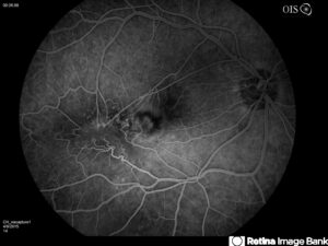
Pupil dilation is essential to diagnose macular pucker, in addition to diagnostic imaging. During your visit, our team will take a “picture” of your retina. This image, called an optical coherence tomography (OCT) scan, can help to actually visualize how much the pucker is pulling on the macula. Furthermore, additional testing with a fluorescein angiogram (FA) may be recommended to help evaluate whether there is leakage in the retina associated with the gliotic or scar tissue. The Figures below show what your retina specialist might see.
 |
 |
How is Macular Pucker Treated?
As retinal specialists, we aim to evaluate various aspects of your vision and eye health to understand how much the macular pucker is contributing to your visual symptoms, and to provide options as to what can be done for you.
Typically, treatment is not indicated unless the patient is significantly affected by symptoms of the pucker. In fact, a small percentage of patients may have spontaneous resolution of this condition, as the membrane has been known to spontaneously retract from the retinal surface.
However, surgery remains a consideration for patients who’s visual complaints are disturbing and create difficulty for them in their daily lives; or for patients who have significant leakage noted on FA imaging, (as the leakage could cause a risk to the patient’s vision over time.)
Retinal surgery is typically performed in an ambulatory setting under local anesthesia, although general anesthesia can be an option. The surgical procedure entails a vitrectomy, (where the vitreous gel is removed from the eye,) and peeling of the membrane from the retinal surface, using specialized instruments.
It may take up to 2-3 months to gain back the majority of vision after the surgery. However, patients who have had macular puckers for extended periods of time or have significant leakage may take longer for recovery.
Do you have questions regarding macular pucker? Call us today! (561) 499-8830
Learn more about Macular Puckers:
The American Society of Retina Specialists. Epiretinal Membranes.
American Academy of Ophthalmology. What is a Macular Pucker?

