What is Age-Related Macular Degeneration?
Age-Related Macular Degeneration (AMD) is a progressive eye condition that affects the macula, the central part of the retina responsible for sharp, detailed vision needed for activities such as reading, driving, and recognizing faces. AMD is one of the leading causes of vision loss in people over the age of 50.
There are two main types of AMD:
-
- Dry AMD: This is the more common form, accounting for about 80-90% of cases. It involves the gradual breakdown of macular cells, leading to thinning and atrophy of the macula. This stage is typically characterized by the accumulation of drusen, small yellow deposits under the retina.
- Wet AMD: Although less common, wet AMD is more severe and leads to more rapid vision loss. It is caused by the growth of abnormal blood vessels beneath the retina that can leak fluid or blood, causing swelling and damage to the macula.
How is a Macular Degeneration Detected?
AMD may develop gradually, making early detection crucial for effective management. An ophthalmologist can diagnose AMD through a series of tests, including:
-
- Dilated Retinal Examination: Eye drops are used to widen the pupils, allowing the doctor to examine the retina and macula for signs of AMD.
- Fundus Photography: This imaging technique captures detailed photographs of the retina, helping to monitor the progression of AMD.
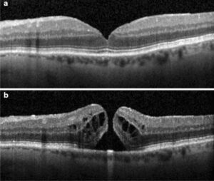
- Optical Coherence Tomography (OCT): OCT provides cross-sectional images of the retina, showing detailed layers and any abnormalities such as fluid accumulation or retinal thinning.
- Fluorescein Angiography (FA): A dye is injected into the bloodstream, and photographs of the retina are taken to visualize the blood vessels and detect any leakage or abnormal growths.
 |
How is Macular Degeneration Treated?
Treatment for AMD depends on the type and severity of the condition:
- Dry AMD: There is currently no cure for dry AMD, but certain treatments can slow its progression:
- Nutritional Supplements: High-dose antioxidants and zinc, as recommended by the Age-Related Eye Disease Study (AREDS), can help slow the progression of dry AMD.
- Lifestyle Modifications: Eating a balanced diet rich in leafy greens and omega-3 fatty acids, along with quitting smoking and managing other health conditions, can contribute to overall eye health.
- Wet AMD: Treatments for wet AMD aim to reduce vision loss and involve:
- Anti-VEGF Injections: Medications injected into the vitreous of the eye can help block the growth of abnormal blood vessels and reduce leakage.
- Laser Therapy: High-energy laser beams are used to destroy abnormal blood vessels and reduce leakage.
- Photodynamic Therapy: A light-sensitive drug is injected into the bloodstream and activated by a laser to destroy abnormal blood vessels.
How is Macular Degeneration Treated?
While there is no cure for AMD, managing the condition effectively can help preserve vision and improve quality of life. Important steps include:
-
- Regular Eye Exams: Frequent monitoring by an ophthalmologist is essential to track the progression of AMD and adjust treatments as needed.
- Healthy Lifestyle: A diet rich in antioxidants, maintaining a healthy weight, and avoiding smoking can help support eye health.
- Low Vision Aids: Devices such as magnifiers, special reading glasses, and electronic tools can assist with daily activities and enhance remaining vision.
If you experience any changes in your vision, such as blurry spots or difficulty seeing fine details, it is important to consult with an eye care professional promptly.
For more information about AMD or to schedule an eye exam, call us today at (561) 499-8830!
