What is a Macular Hole?
A macular hole is a central defect in the retina. The macula is the central region of the retina, and is responsible for critical vision including reading, driving, and watching television.
A hole can develop as the vitreous gel ages. The vitreous gel goes through changes with age, which frequently results in floaters. Abnormal adherence of the vitreous to the macular region may result in traction or pulling, resulting in a macular hole.
How is a Macular Hole Detected?
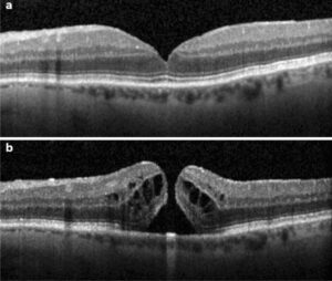
There are 4 stages of a macular hole, all of which can result in some degree of symptoms. The most common symptom includes distortion and reduced vision for both near and distance. A macular hole is diagnosed by the ophthalmologist after dilated retinal examination. Optical coherence tomography (OCT) helps evaluate macular holes.
 |
How are Macular Holes Treated?
Refinements in the surgical treatment of macular hole have steadily improved outcomes. The procedure involves the removal of the vitreous gel (vitrectomy), placement of a temporary gas bubble in the eye, and often removal of a membrane surrounding the hole.
Surgery is typically performed in an ambulatory setting under local anesthesia, although general anesthesia can be utilized. The gas bubble is put in the eye so the hole can close under a “dry” environment; the fluid in the eye can keep the hole open.
To keep fluid away from the hole while it closes, it is necessary for the patient to remain in a prone (face down) position anywhere from several days to 2 weeks. It may take up to 6 weeks for the air bubble to completely resolve from the eye.
Patients with macular holes may have varying degrees of visual loss. It is possible that, if not operated on, the condition may still remain stable with no further loss in vision. However, there also may be deterioration over time. This is impossible to predict.
Visual results after surgery depend on a number of factors including the presence of a cataract, the longevity of the hole, and any other associated macular conditions such as macular degeneration. Typically, it has been shown that if a hole is present for less than 6 months, at least a 3-line improvement in vision will occur with hole closure.
It can take anywhere from 2 to 3 months to regain vision after surgery. Vision is often diminished from cataract progression, and maximal vision improvement may require cataract surgery. Approximately 5-10% of macular holes are not closed successfully with surgery. Sometimes repeat surgery may lead to closure.
Do you have questions regarding macular holes? Call us today! (561) 499-8830
