What is Optical Coherence Tomography?
Optical Coherence Tomography scans, or OCTs, are a quick and non-invasive test used to capture cross-section images of the retina, allowing for details of the retinal layers to be visualized and measured. OCTs help your provider to further understand your current retinal condition and to determine if, and what type, of treatment might be needed.
What Conditions Can be Diagnosed with OCT?
The OCT is the cornerstone of our office due to the detailed images it is able to produce. The following includes common conditions that are able to be diagnosed with OCT:
-
- Diabetic Retinopathy: OCT can reveal changes in the retina caused by diabetes, such as fluid leakage, retinal thickening, and damage to the retinal blood vessels.
- Age-Related Macular Degeneration (AMD): Helps detect drusen (yellow deposits) and other changes in the macula, such as fluid accumulation or neovascularization (abnormal blood vessel growth).
- Macular Edema: Assesses the extent of fluid buildup in the macula, which can affect central vision.
- Glaucoma: Provides detailed images of the optic nerve head and retinal nerve fiber layer, helping to detect and monitor glaucoma-related damage.
- Retinal Detachment: Identifies signs of retinal separation from the underlying layers and helps in assessing the extent of the detachment.
- Retinal Vein and Artery Occlusions: Detects swelling and other changes in the retina resulting from blood vessel blockages.
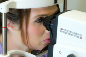
How is Optical Coherence Tomography Performed?
The OCT procedure is quick, comfortable, and involves the following steps:
-
- Preparation: You will be asked to sit in front of the OCT machine and place your chin on a rest to keep your head steady.
- Imaging: You will be instructed to look at a fixed point while the machine scans your eye. The OCT device uses a beam of light to capture cross-sectional images of the retina.
- Image Analysis: The obtained images are analyzed by the ophthalmologist to assess the retinal structures and identify any abnormalities.
Benefits of Optical Coherence Tomography
-
- High Resolution: Provides detailed, high-resolution images of the retinal layers and optic nerve, allowing for precise diagnosis and treatment planning.
- Non-Invasive: The procedure is painless and does not involve any direct contact with the eye, making it comfortable for patients.
- Early Detection: Facilitates early detection of retinal and optic nerve conditions, which is crucial for timely intervention and effective management.
- Monitoring: Useful for tracking the progression of diseases and the effectiveness of treatments over time.
What to Expect After the Procedure
-
- No Downtime: There is no recovery time needed, and you can resume normal activities immediately after the OCT scan.
- Results: The results of the OCT scan will be reviewed by your ophthalmologist, who will discuss the findings and recommend any necessary follow-up or treatment based on the images.
For more information or to schedule an eye exam, contact us today at (561) 499-8830!
Learn more about Retinal Tears/Detachments:
Cleveland Clinic. Optical Coherence Tomography.
American Academy of Ophthalmology. What is Optical Coherence Tomography?
